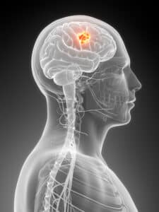
Learn everything you need to know about Brain Tumor Treatment with dedicated neurosurgeons in the areas of Dallas, TX
What is Craniotomy Procedure (Brain Tumor Treatment)?
Craniotomy is a surgical procedure in which part of the skull is removed in order to view the brain. The piece of skull removed is called a “bone flap.” After the surgery is performed to remove the brain tumor, the bone flap is fitted back into the skull.
Surgery is often the most effective way to treat many brain tumors, whether they are benign or malignant. Craniotomies, which are performed by our expert team of neurosurgeons in our Dallas offices, are designated in different ways. A frontotemporal, parietal, temporal or suboccipital craniotomy is named after the bone that is removed.
In order to accurately access the tumor and reduce the risk of damage to healthy brain tissue, imaging devices may be used to help guide the surgeon. Known as a stereotactic craniotomy, scans are taken that create a three-dimensional image of the brain. A computer provides a navigation system to safely route the surgeon to the precise location of the tumor.

The Craniotomy Procedure
A craniotomy or brain tumor surgery is performed, sometimes under general anesthesia, in a hospital. An incision is made in the scalp (which is shaved at the incision site), and the bone flap is removed to allow access to the treatment area. There are cases in which patients remain awake during surgery, and are asked to move their legs, recite the alphabet or tell stories to ascertain whether brain functioning has been affected.
The location of the incision will depend upon the area being treated. A medical drill may be used to create small holes, or a special saw may be used to cut the bone flap. An incision is then made in the dura matter, which is the covering of the brain. The tumor is located and removed in its entirety if possible. Once the procedure is complete, any tissue that has been cut into is stitched together, and the bone flap is reattached using plates, sutures or wires.
Depending on the location of the tumor, complete removal may be difficult. However, even if the entire tumor cannot be removed, surgery can relieve symptoms and help reduce pressure within the brain. If the tumor has been only partially removed, additional treatment, such as radiation therapy, may be required to destroy any remaining tissue.

Risks of Craniotomy
Surgery to treat a brain tumor is complex, and not without risks and potential complications. Some risks are related to the specific location being operated on. If, for example, the area of the brain being operated on controls speech, then speech may be affected. Aside from the usual surgical risks of infection, bleeding and the formation of blood clots, other risks specific to craniotomy include swelling and accumulation of fluid, which can lead to brain damage and other serious complications. Surgery can also damage healthy brain tissue, causing problems with thinking, seeing and speaking.
Recovery from Brain Tumor Treatment
Post-craniotomy, a patient usually has a headache for a few days, and can feel tired or weak. Most patients spend between 3 and 7 days in the hospital, but specific recovery times vary. Pain medication is prescribed in order to relieve symptoms and promote healing. Restrictions will be placed on driving, lifting and many common daily activities until the patient is cleared by the surgeon.
If the brain tumor is determined to be malignant during the craniotomy, additional treatment may be necessary. Treatment may include radiation therapy, which uses high energy X-rays or gamma rays to destroy malignant cells, or chemotherapy, which involves the use of drugs administered intravenously.
Schedule Your Consultation Today!
Call 214-823-2052 to schedule your consultation with one of our providers today!

